
Anti-IDH1 R132H (Hu) from Mouse (H09) – unconj. – prediluted RTU


-
Overview
SKU DIA-H09-L Specificity Species Reactivity Immunogen Host Species Isotype Clone Clonality (Mono-/Polyclonal) Application Immunohistochemistry (IHC), Immunohistochemistry (Paraffin-embedded Sections)
Conjugation Dilution Format in Antibody Diluent Buffer, purified prediluted antibody, Ready-to-use
Application Note Product line / Topic Intended Use Temperature - Storage Temperature - Transport Search Code Manufacturer / Brand Uniprot_ID Gene_ID Alias Cytosolic NADP-isocitrate dehydrogenase, IDH1, IDP, Isocitrate dehydrogenase [NADP] (cytoplasmic), Isocitrate Dehydrogenase 1, Soluble, NADP(+)-specific ICDH, Oxalosuccinate decarboxylase, PICD
- Datasheets and Downloads
-
Additional Product Information
Since 2009 clone H09 (Heidelberg 2009) has established worldwide as a standard tool for IHC detection of IDH1 R132H in neuropathology and research. This is documented by a high number of publications as cited via CiteAB. Anti-IDH1 R132H antibody clone H09 (exclusively licenced from the German Cancer Research Center by dianova) is available in different formats:
Cat.No. Product Form Status Qty Price DIA-H09 anti-IDH1 R132H, clone H09 concentrated, lyophilized CE IVD 500 µl 482,00 € DIA-H09-M anti-IDH1 R132H, clone H09 concentrated, lyophilized CE IVD 100 µl 142,00 € DIA-H09-L anti-IDH1 R132H, clone H09 prediluted, ready-to-use RUO 8 ml 482,00 € Immunohistochemistry of human IDH1 R132H in formalin-fixed paraffin-embedded brain tissue sections
A
B
C
D
E
F
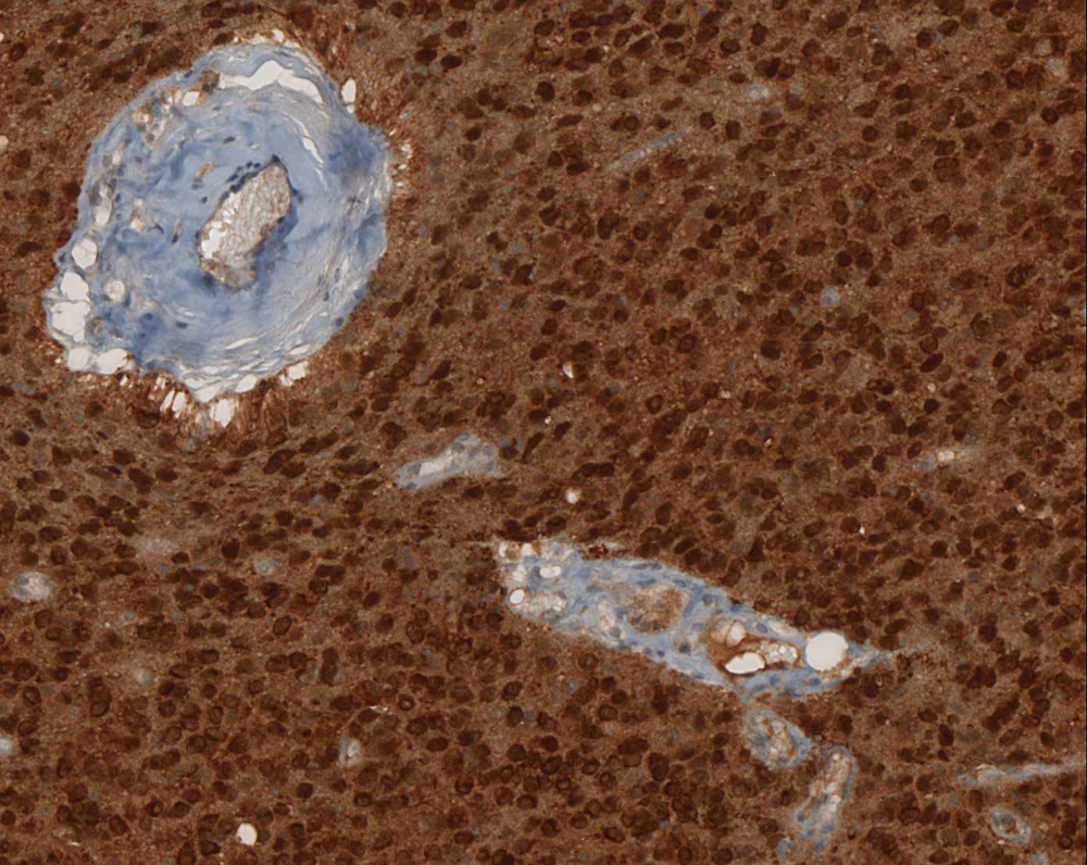
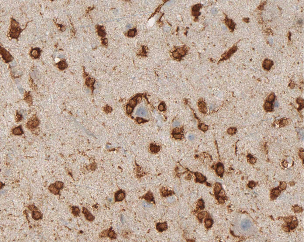
A: Strong reaction of IDH1 mutation specific antibody clone H09 in tumor center of anaplastic oligoastrocytoma.
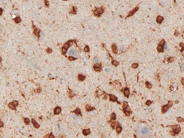
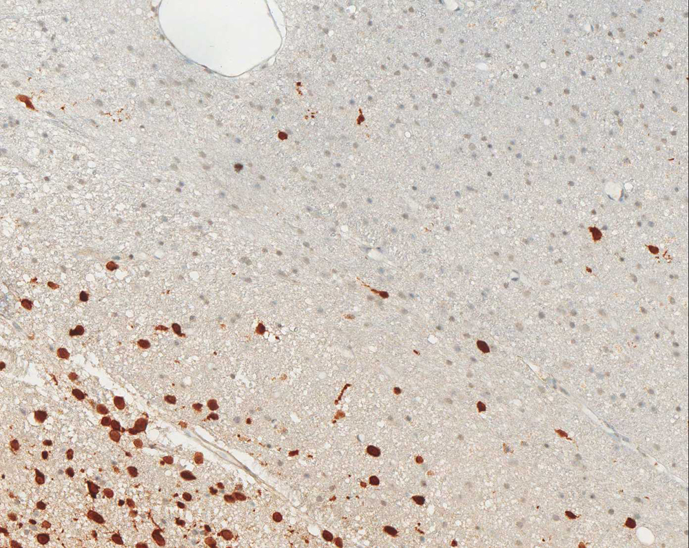
B: Infiltration zone of anaplastic astrocytoma with specific labelling of infiltrating glioma cells by antibody clone H09.
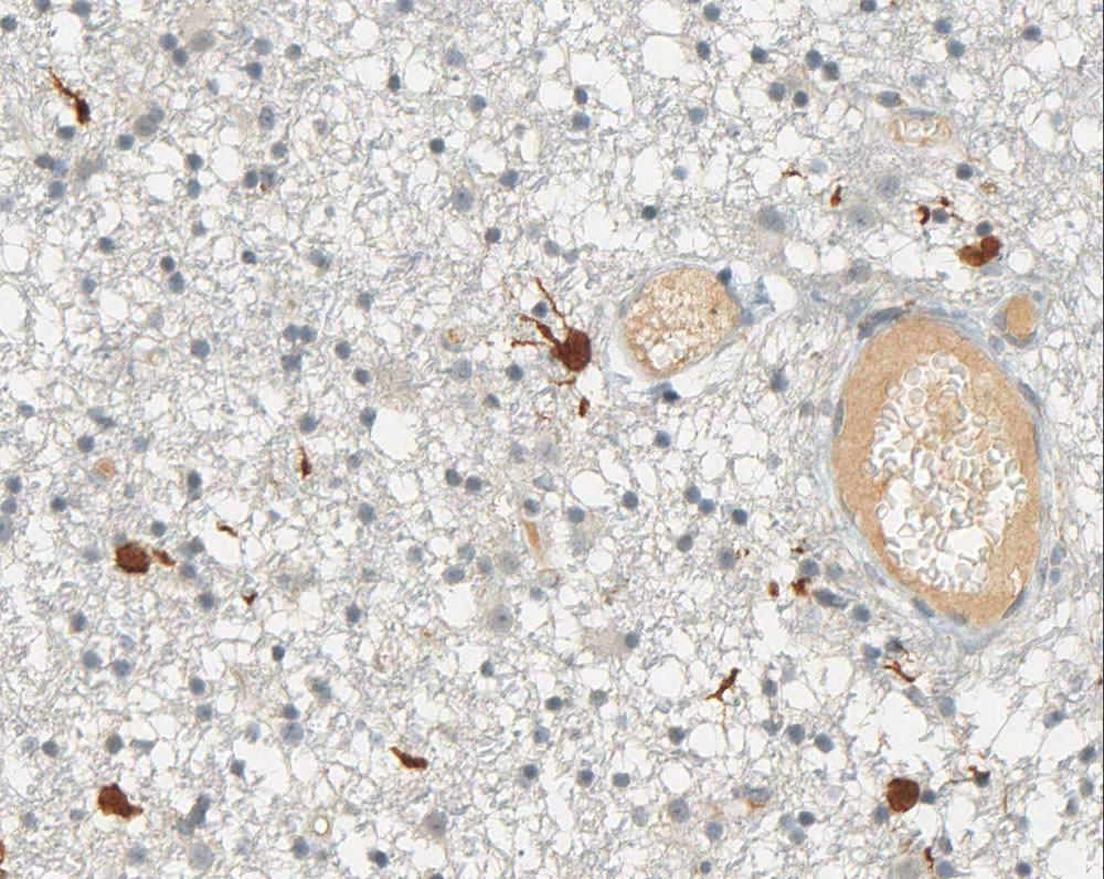
C: Identification of single tumor cells in white matter distant from tumor center with antibody clone H09.
D: Cortex infiltrated by oligodendroglioma with specific labelling of tumor cells by antibody clone H09.
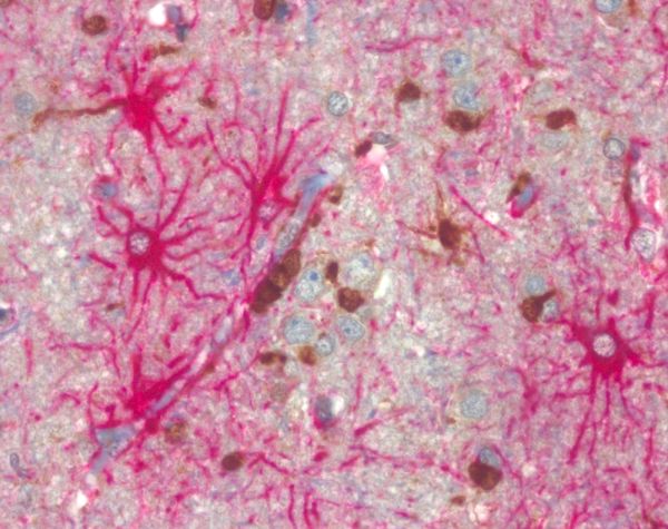
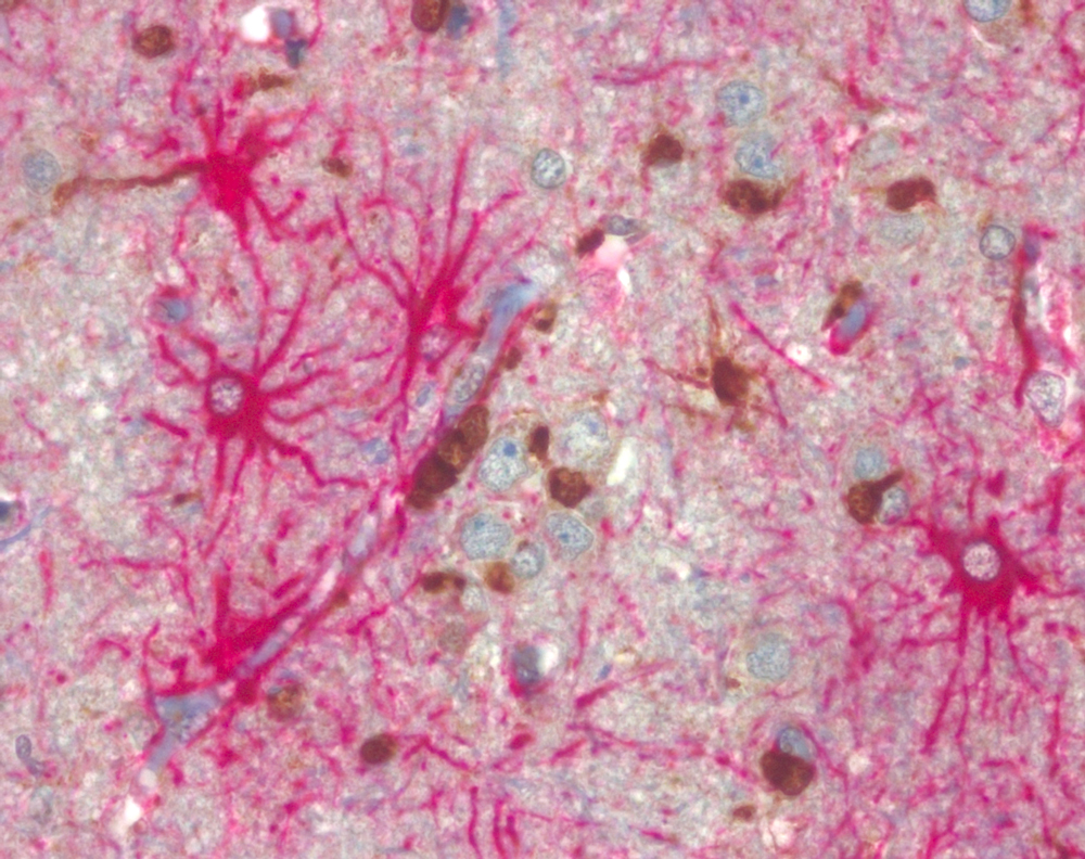
E: Double staining of GFAP (glial fibrillary acidic protein, red) and clone H09 (brown) of oligodendroglioma infiltration zone demonstrating specific labelling of tumor cells but not of GFAP positive reactive astrocytes.
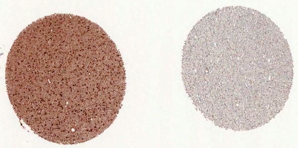
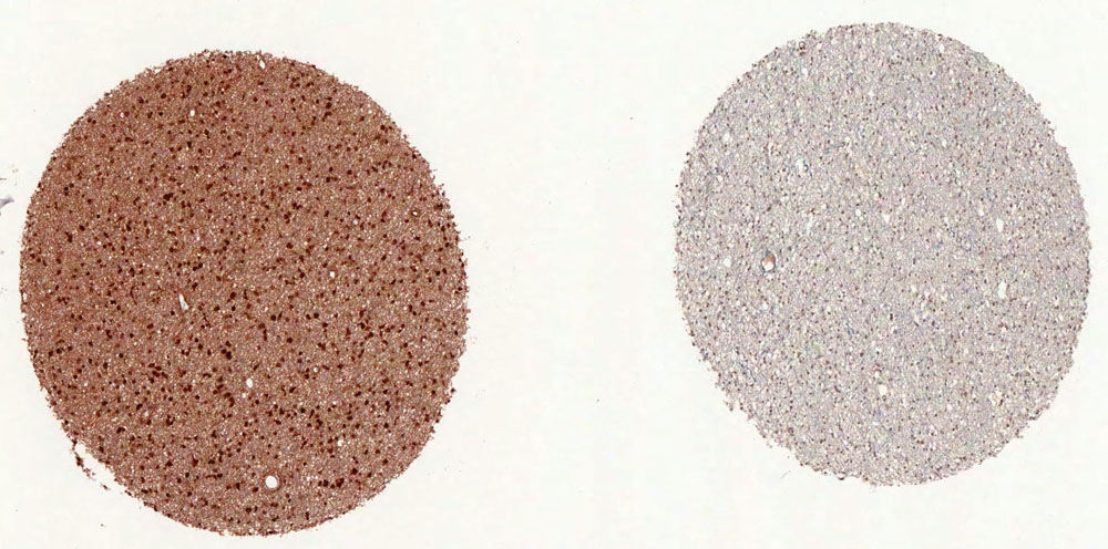
F: Strong reaction of antibody clone H09 with IDH1 R132H mutated diffuse astrocytoma (left) but not with wild type tumor (right).(pictures courtesy of Prof. Dr. med. Andreas von Deimling, Department of Neuropathology, University Heidelberg/
Clinical Co-operation Unit Neuropathology, German Cancer Research Center (DKFZ), Heidelberg, Germany)Background
Antibody clone H09 reacts specifically with the isocitrate dehydrogenase 1 (IDH1) R132H point mutation in tissue sections from formalin-fixed brain tumor. Heterozygous point mutations of IDH1 codon 132 are frequent in World Health Organization (WHO) grade II and III gliomas. IDH1 R132H mutations occur in approximately 70% of astrocytomas and oligodendroglial tumors. The high frequency and distribution of the IDH1 R132H mutation among specific brain tumor entities allow the highly sensitive and specific discrimination of various tumors by immunohistochemistry, such as anaplastic astrocytoma from primary glioblastoma or diffuse astrocytoma WHO grade II from pilocytic astrocytoma or ependymoma. Noteworthy is the discrimination of the infiltrating edge of tumors with IDH1 mutation from reactive gliosis.
Combined immunohistochemistry of ATRX (dianova clone AX1) and IDH1R132H (dianova clone H09)
substitutes molecular testing.The routine practical approach for diagnosing astrocytomas and oligodendrogliomas begins with perfoming IHC for IDH1 R132H and ATRX expression. Stepwise analysis of molecular parameters with initial IHC for ATRX and IDH1 R132H followed by 1p/19q analysis and then by IDH sequencing significantly reduces the number of molecular tests required for unequivocal diagnosis (Reuss et al., 2015). Learn more…
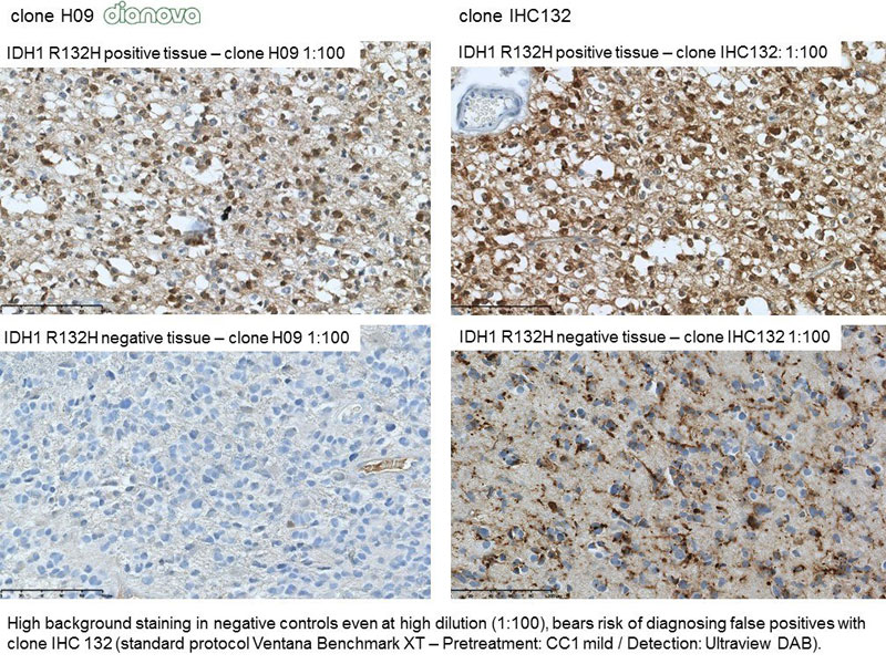
Comparative study
References
- Capper D et al. Monoclonal antibody specific for IDH1 R132H mutation. Acta Neuropathol. 118(5): 599-601, 2009
- Capper D et al. Characterization of R132H mutation-specific IDH1 antibody binding in brain tumors. Brain Pathol. 20(1): 245-254, 2010
- Preusser M et al. IDH testing in diagnostic neuropathology: review and practical guideline article invited by the Euro-CNS research committee. Clinical Neuropathology, 30(5):217-230, 2011
- Van den Bent MJ et al. Interlaboratory comparison of IDH mutation detection. J Neurooncol 112:173–178, 2013
- Schumacher T et al. A vaccine targeting mutant IDH1 induces antitumour immunity. Nature 2014, DOI:10.1038/nature13387
- Reuss DE et al. ATRX and IDH1-R132H immunohistochemistry with subsequent copy number analysis and IDH sequencing as a basis for an „integrated“ diagnostic approach for adult astrocytoma, oligodendroglioma and glioblastoma. Acta Neuropathol. 129(1):133-146, 2015
- David NL et al. The 2016 World Health Organization Classification of Tumors of the Central Nervous System: a summary. Acta Neuropathol., 131:803-820, 2016
-
Images
IDH1 R132H: infiltration zone glioma IDH1 R132H: Cortex infiltrated by oligodendroglioma IDH1 R132H: Double staining of GFP and clone H09 IDH1 R132H: Identification of single tumor cells IDH1 R132H: infiltrating glioma cells IDH1 R132H: strong reaction on mutated diffuse astrocytoma IDH1 R132H: tumor center of anaplastic oligoastrocytoma.