Anti-Fibroblasts (CD90, Thy-1) (Hu) from Mouse (AS02) – unconj.


-
Overview
SKU DIA-100 Specificity Species Reactivity Immunogen Host Species Isotype Clone Clonality (Mono-/Polyclonal) Application Flow Cytometry, Immunofluorescence, Immunohistochemistry (frozen sections), Immunohistochemistry (IHC), Immunoprecipitation, Western Blot
Conjugation Dilution Cell Separation 1:50 – 1:200, Flow Cytometry 1:50 – 1:200, Immunofluorescence 1:50 – 1:200, Immunohistochemistry (IHC): 1:50 – 1:200, Western Blot (WB), non-reducing: 5 – 10 ng/ml
Format 0.05% NaN3, 2% BSA, in PBS (pH 7.4), lyophilisate, ProteinA/G purified antibody (from culture supernatant)
Product line / Topic Intended Use Temperature - Storage Temperature - Transport Search Code Manufacturer / Brand Uniprot_ID Gene_ID Alias CD90, CDw90, Thy-1 antigen, Thy-1 membrane glycoprotein, THY1
- Datasheets and Downloads
-
Additional Product Information

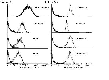
Flow Cytometry – Anti-CD90 / Thy1 antibody (DIA-100, DIA-120). The antibody strongly reacts with human dermal fibroblasts whereas it does not stain lymphocytes, keratinocytes, monocytes, granulocytes, thrombocytes, HUVEC, and HDMC. Monoclonal antibody AS02 (▬▬▬) compared to an appropriate cell marker (——–).

Western blot – Anti-CD90 / Thy1 antibody (DIA-100). Detection of CD90 (Thy-1) protein in cell extract of human fibroblasts by Western blotting with 0.6 µg/ml anti-CD90 / Thy1 antibody clone AS02 (DIA-100). CD90 / Thy-1 detection by the antibody AS02 shows a band at 30-32 kDa under reducing and nonreducing conditions, with better reactivity using nonreduced sample. The epitope bound by AS02 resides within the 15-kDa polypeptide chain, and not in the N-linked oligosaccharide side chains or the GPI anchor structure.

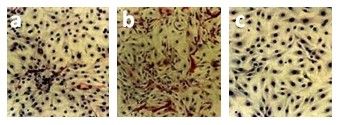
Immunohistochemistry – Anti-CD90 / Thy1 antibody (DIA-100). Thyrocyte cell culture before (a, b) and after (c) the elimination of contaminating fibroblasts using anti-CD90 antibody clone AS02 (DIA-100) coupled to magnetic beads. Immunostaining of Fibroblasts (red) with AS02, nuclei counterstained with haemalaun.
a) Thyrocyte culture after the first passage; single fibroblasts (red) are visible
b) Thyrocyte culture after the fourth passage; many fibroblasts (red) are visible
c) Thyrocyte culture after elimination of fibroblasts using AS02 magnetic beads (fifth passage).
Immunofluorescence / Immunocytochemistry – Anti-CD90 / Thy1 antibody (DIA-100, DIA-120). Immunofluorescence double staining of cultured human dermal fibroblasts with anti-CD90 / Thy1 antibody clone AS02 (green) and anti-prolylhydroxylase antibody (yellow). (magnification x 300)
-
Images

00982_Abb1_Flow-Cytometry-Reactivity-of-anti-CD90-clone-AS02 
00982_Abb2_IF-staining-of-fibroblasts-with-anti-CD90-clone-AS02 
00982_Abb3_Thyrocyte-culture-before-and-after-elimination-of-fibroblasts-with-anti-CD90-DIA-100 
00982_Abb4_WB-anti-CD90-clone-AS02-DIA-100
