
1. Controls
In order to distinguish specific from non-specific bands of the sample material, always run a control on every blot, in which only secondary antibody conjugate without primary antibody is applied.
2. Dilution of secondary antibody
The secondary antibody should not be used at too high concentrations. In most applications the secondary antibody can be used in dilutions well above 1:10.00 while higher concentrations may favor nonspecific background binding. As a guideline, in comparison to color substrates the secondary antibody dilution can be doubled when ECL-Substrates are used. The optimal dilution may also vary with different batches of secondary antibody.
Recommended dilutions for Western blotting:
• HRPO conjugates: 1:5.000 – 1:100.000 (non-ECL) / 1:10.000 – 1:200.000 (ECL)
• Alk. Phos. conjugates: 1:5.000 – 1:50.000
• Biotin conjugates: 1:20.000 – 1:400.000
• Streptavidin-HRPO: 0,01 – 0,1 µg/ml
• Streptavidin-Alk.Phos.: 1 – 2 µg/ml
3. Attention when using bovine milk powder
Bovine milk powder is frequently used as blocking reagent in Western blotting, but sometimes it can also be the source of unwanted background. In order to reduce the background reaction of the secondary antibody with bovine Ig in milk powder the use of secondary antibodies adsorbed against bovine (MinX Bo) or human (MinX Hu) Ig / serum proteins is recommended.
Other measures to reduce non-specific background include blocking with 5% normal serum from the same species as the secondary antibody or with IgG-free BSA (Cat. No. 001-000-161 / 2). Often, switching to normal serum as blocking reagent for problematic blots efficiently leads to cleanest results.
Generally, the secondary antibody should be diluted in buffer without protein base such as milk powder a.o. in order to exclude reactions of the secondary antibody with immunoglobulins or proteins present in the protein supplement. This is particularly important for anti-goat conjugates (see 4.).
4. Attention when using anti-goat conjugates
Anti-goat secondary antibodies from the host species donkey, rabbit, and mouse highly cross-react to bovine immunoglobulins, for example in milk powder. If the secondary antibody dilution buffer contains milk powder, almost all of the anti-goat conjugate will be adsorbed by bovine Ig. This leads to either a complete lack of signal or a faint signal, if the conjugate is applied at high concentrations. Additionally these conjugates may produce background on the blot, if bovine milk powder was used for blocking.
For the detection of goat primary antibodies on Western blots, we recommend anti-goat from bovine (cat. 805-xx5-180). Production of anti-goat antiserum in the very closely related host bovine results in antibodies with no cross-reactivity to bovine Ig / serum proteins.
5. Endogenous immunoglobulins in sample lysate
If the sample material separated by SDS-PAGE is a lysate of immunoglobulin-containing cells or tissues, secondary antibodies without cross-reaction (MinX) to the same species than the sample should be used. (Alternatively, normal serum (Ig) of the same species than the sample material can be added to the secondary antibody dilution buffer.)
6. Background bands from antibodies after immunoprecipitation
In the following article you can read how background bands caused by precipitating immunoglobulins on the blot can be avoided when using secondary antibodies for Western blot detection.
Immunoblot (Western Blot) – Avoiding background bands from antibodies
One of the most common problems in immunoblotting (Western blot) using secondary antibodies for indirect detection are background bands caused by antibodies in sample preparations originating from, e.g. immunoprecipitation, cell-or tissue-own immunoglobulins in the lysate or contamination with bovine immunoglobulins (FCS in cell lysates, BSA, etc.). The antibody heavy and light chains slotted from the sample after reducing SDS-PAGE display bands at 50 kDa and 25 kDa, respectively. Under non-reducing conditions gamma immunoglobulins (IgG) show a band at approximately 150 kDa.
Paying attention to this issue is most important in immunoprecipitations (IP); especially after immunoprecipitation when the target protein is expected in the range of 25kDa or 50kDa this issue must be addressed.
The problem of antibody background bands after IP can completely be bypassed by using directly conjugated primary antibodies for the detection of the target protein. However, labeled primary antibody is often not available, so that an unconjugated primary must be detected with a secondary antibody conjugate.
One possible way to prevent antibodies from being blotted after immunoprecipitation includes crosslinking antibody to Protein A/G-coated beads or covalently coupling antibody directly to treated beads. However, in order to avoid antibody contamination using this approach, eluting the antigen under non-denaturing conditions is crucial, as otherwise the denatured antibody fragments will be eluted with the antigen.
If the antigen on the blot is indirectly detected using unconjugated primary antibody and a labeled secondary antibody, antibody bands can be avoided by chosing the right secondary antibody under certain requirements:
The primary antibodies used for immunoprecipitation and detection on the blot:
1. are from different host species.
Background bands of the precipitating antibody on the blot can be avoided by secondary antibodies without cross-reaction (MinX) to the species of the precipitating antibody.
Example: Precipitation with polyclonal rabbit anti-target protein, detection on blot with monoclonal mouse anti-target protein
> Secondary antibody anti-mouse IgG (H+L) MinX Rabbit etc. (e.g. cat. 115-035-146).
List of anti-IgG/M (H+L) without inter-species cross-reaction for Western Blots
Find more anti-Ig(H+L) specific antibodies for Western blots at: Secondary Antibodies
2. are from the same host species and
a. have different isotypes.
By
the selection of a secondary antibody that is specific for the
sub/class of the primary detecting antibody the recognition of the
precipitating antibody on the blot can be avoided.
Example:
Precipitation with monoclonal mouse (IgG1) anti-target protein,
detection on blot with monoclonal mouse (IgG2a) anti-target protein
> Secondary antibody anti-mouse IgG2a (e.g. cat. 115-035-206).
List of Anti-IgG subclass and Anti-IgM specific secondary antibodies for Western blots
b. the target protein is near 50 kDa.
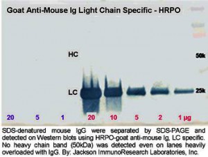
Secondary antibodies against the light chains of mouse IgG do not react with the reduced and denatured heavy chain (50 kDa) of the precipitating antibody. They only bind to the reduced, denatured light chain of the precipitated antibody on the blot (see Figure) and to the native light chains of the primary antibody used for detection. Therefore, a 50 kDa protein may be detected on blots without interference from the precipitating antibody. Only one antibody band in the region of the light chains, but not in the range of the target protein is detected.
Example:
Target protein approx. 50 kDa; precipitation using monoclonal mouse
(IgG1) anti-target protein, detection on blot with monoclonal mouse
(IgG1) anti-target protein
> Secondary antibody anti-mouse Ig/G light chain (e.g. cat. 115-035-174).
c. the target protein is near 25 kDa.
Secondary antibodies against the Fc fragment of mouse IgG do not react with the reduced and denatured light chain (25 kDa) of the precipitating antibody. They only bind to the reduced, denatured heavy chain of the precipitated antibody on the blot and to heavy chains of the native primary antibody used for detection. Therefore, a 25 kDa protein may be detected on blots without interference from the precipitating antibody. Only one antibody band in the region of the heavy chains, but not in the range of the target protein is labeled.
Example: Target protein approx. 25 kDa; precipitation using monoclonal mouse (IgG1) anti-target protein, detection on blot with monoclonal mouse (IgG1) anti-target protein
> Secondary antibody anti-mouse IgG Fc (e.g. cat. 115-035-071).
List of anti-IgG Fc specific secondary antibodies for Western blots
Note: If the primary antibody recognizes its target protein in non-reduced form and if the target protein related to its size etc. can be displayed in the SDS-PAGE, the sample may be applied without reducing reagents. The primary antibodies used to precipitate then forms only one antibody band at 150 kDa which does not interfere with the detection of target proteins outside this molecular weight range.
Anti-Ig/G Light Chain Specific Secondary Antibodies
Anti-Ig/G light chain specific antibodies react with native primary antibodies used for the detection of proteins in Western blots. If applied in an appropriate dilution they do not bind to the reduced and denatured IgG heavy chain band (50 kDa) on blots. This makes them ideal for the indirect detection of antigens near 50 kDa after immunoprecipitation, where large amounts of reduced and denatured IgG heavy chain can obscure the detection of the target protein.
Although the anti-light chain specific antibodies show a strong reactivity with native IgG light chains in Western blot, the reduced and denatured form on the blot may not be recognized as sensitive. We therefore do not recommend using the antibodies for a sensitive / quantitative detection of IgG light chains by Western blotting.
In order to prevent cross-reactions to immunoglobulins from other species which may be present on blots, the antibodies have been adsorbed to serum proteins from many other species.CopyPDFShow entriesSearch:
| Cat. No. | Product | Size | IgG type |
|---|---|---|---|
| 112-035-175 | Goat anti-Rat Ig/G, light chain-HRPO, MinX Bo,Go,Ho,Hu,Rb,Rt,Sh | 0,5 ml | IgG |
| 112-055-175 | Goat anti-Rat Ig/G, light chain-Alk. Phos., MinX Bo,Go,Ho,Hu,Rb,Rt,Sh | 0,5 ml | IgG |
| 112-065-175 | Goat anti-Rat Ig/G, light chain-Biotin, MinX Bo,Go,Ho,Hu,Rb,Rt,Sh | 0,5 ml | IgG |
| 115-035-174 | Goat anti-Mouse Ig/G, light chain-HRPO, MinX Bo,Go,Ho,Hu,Rb,Rt,Sh | 0,5 ml | IgG |
| 115-055-174 | Goat anti-Mouse Ig/G, light chain-Alk. Phos., MinX Bo,Go,Ho,Hu,Rb,Rt,Sh | 0,5 ml | IgG |
| 115-065-174 | Goat anti-Mouse Ig/G, light chain-Biotin, MinX Bo,Go,Ho,Hu,Rb,Rt,Sh | 0,5 ml | IgG |
| 205-032-176 | Mouse anti-Goat Ig/G, light chain-HRPO, MinX Ho,Hu,Ms,Rb,Rt | 0,5 mg | IgG |
| 205-052-176 | Mouse anti-Goat Ig/G, light chain-Alk. Phos., MinX Ho,Hu,Ms,Rb,Rt | 0,5 mg | IgG |
| 205-062-176 | Mouse anti-Goat Ig/G, light chain-Biotin, MinX Ho,Hu,Ms,Rb,Rt | 0,5 mg | IgG |
| 211-032-171 | Mouse anti-Rabbit Ig/G, light chain-HRPO, MinX Bo,Go,Ha,Ho,Hu,Ms,Rt,Sh | 0,5 mg | IgG |
Showing 1 to 10 of 15 entriesFirstPrevious12NextLast
Target proteins near 25 kDa may be detected indirectly after immunoprecipitation without any interference in this range by using anti-IgG Fc fragment specific antibodies, because they do not react with the reduced and denatured antibody light chains on the blot.
Figures:
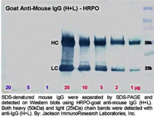
Fig. 1. Lanes 1, 2: detection of primary antibodies made in goat against a 50 kDa target protein; Lanes 3, 4: detection of heavy (50 kDa) and light (25 kDa) chains of reduced and SDS-denatured goat IgG separated by SDS-PAGE; Lanes 5, 6: reduced and SDS-denatured mouse IgG (control).
Fig. 2. Lanes 1-3: reduced and SDS-denatured mouse IgG (control); Lanes 4-8: detection of heavy (50 kDa) and light (25 kDa) chains of reduced and SDS-denatured goat IgG separated by SDS-PAGE.
Lanes 3, 4 (Fig. 1) and Lanes 4-8 (Fig. 2) demonstrate the relatively weak reactivity of the anti-goat light chain specific antibody with reduced and SDS-denatured light chains.
(By: Jackson ImmunoResearch Laboratories, Inc.)
Fluorescent-labelled Secondary Antibodies for Western Blotting
• compatible with LI-COR® Odyssey
• quantitative Western Blotting
• In-Gel Western Blotting
• In-Cell- and On-Cell Western Arrays
• highest Sensitivity and Specificity
Alexa Fluor® 680 and Alexa Fluor® 790
Alexa Fluor® 680 is a far-red-emitting dye with peak excitation at 684nm and peak emission at 702nm. Alexa Fluor® 790 is an infrared-emitting dye with peak excitation at 783nm and peak emission at 803 nm (Figure 1).
Antibodies conjugated with far-red- and Infrared-emitting dyes are more sensitive than those with dyes emitting visible light due to low fluorescence quenching of the conjugates, high extinction coefficients of the dyes, and low background autofluorescence. The increased brightness allows for a wider range of immunofluorescence detection and imaging modalities. Far-red and Infrared dye conjugates can be used for higher sensitivity Western blots, quantitative Western blots, in-gel Western blots, microWestern arrays, in-cell Western arrays, on-cell Western arrays, tissue section imaging, small animal whole body imaging, and other techniques that require the brightest dyes.
Alexa Fluor® 680 and Alexa Fluor® 790 for Far Red- and Infrared Western Blot Detection
| Conjugate | Alexa Fluor 680 | Alexa Fluor 790 |
|---|---|---|
| Streptavidin | 016-620-084 | 016-650-084 |
| Conjugate | Alexa Fluor 680 | Alexa Fluor 790 |
|---|---|---|
| Donkey IgG, whole molecule | 017-620-003 | 017-650-003 |
| Goat IgG, whole molecule | 005-620-003 | 005-650-003 |
| Mouse IgG, whole molecule | 015-620-003 | 015-650-003 |

Fig. 2 :Excitation and emission spectra of Alexa Fluor® 680 (left)- and Alexa Fluor® 790 (right)-conjugated secondary antibodies. All peaks were normalized. Spectra were obtained with a M-Series spectrofluorometer from Photon Technology International, Inc. and an Ultraspec 1100 pro from Amersham Biosciences (Fig. Jackson ImmunoResearch).
Here you will find our Western Blot Troubleshooting Guide (German).