
Goat IgG anti-Mouse IgG1 (Fc)-FITC, MinX Hu
-
Overview
SKU SBA-1070-02 Host Species IgG Form Species Reactivity Specificity Isotype Clonality (Mono-/Polyclonal) Application FLISA, Flow Cytometry, IHC (Whole Mount), Immunocytochemistry, Immunohistochemistry (frozen sections), Immunohistochemistry (Paraffin-embedded Sections), Western Blot
Conjugation Maximum Absorption Maximum Emission No Cross-reactivity (MinX) with Dilution Format Intended Use Manufacturer / Brand - Datasheets and Downloads
-
Images
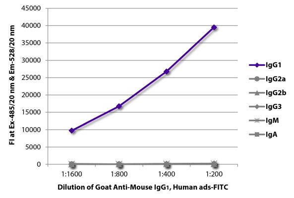
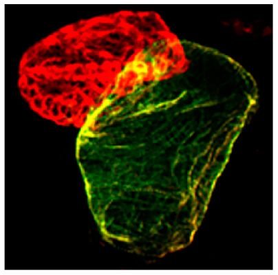
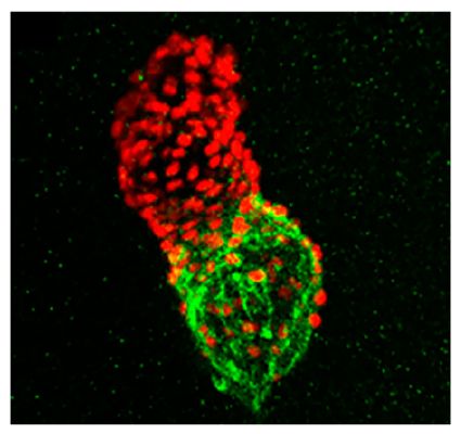
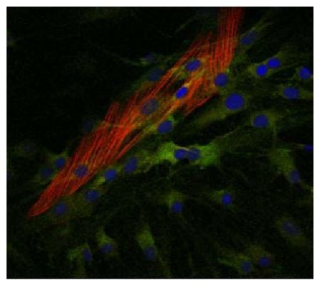
FLISA plate was coated with purified mouse IgG1, IgG2a, IgG2b, IgG3, IgM, and IgA. Immunoglobulins were detected with serially diluted Goat Anti-Mouse IgG1, Human ads-FITC (SB Cat. No. 1070-02). Zebrafish embryos were stained with anti-atrial myosin heavy chain and anti-sacromeric myosin heavy chain followed by Goat Anti-Mouse IgG1, Human ads-FITC (SB Cat. No. 1070-02) and Goat Anti-Mouse IgG2b, Human ads-TRITC (SB Cat. No. 1090-03) secondary ant Tg(myl7:nDSRed) transgenic zebrafish embryos were stained with anti-atrial myosin heavy chain and anti-DsRed followed by anti-rabbit AF594 and Goat Anti-Mouse IgG1, Human ads-FITC (SB Cat. No. 1070-02) secondary antibodies. Image from Zhu D, Fang Y
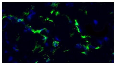
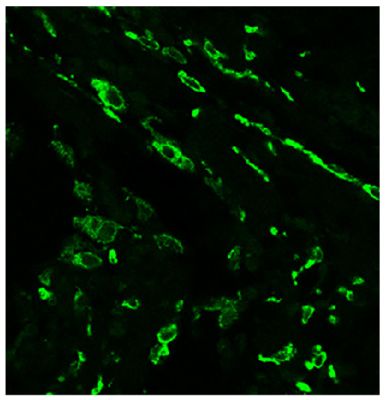
Frozen equine sacroid tissue section was stained with anti-CD4 followed by Goat Anti-Mouse IgG1, Human ads-FITC (SB Cat. No. 1070-02) and DAPI. Image from Wilson AD, Hicks C. Both tumour cells and infiltrating T-cells in equine sarcoids express FOX
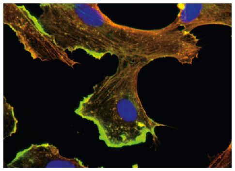
BMH29L cells were stained with anti-β-actin and anti-γ-actin followed by Goat Anti-Mouse IgG1, Human ads-FITC (SB Cat. No. 1070-02) and Goat Anti-Mouse IgG2b, Human ads-TRITC (SB Cat. No. 1090-03) and DAPI. Image from Pasquier E, Tuset M, Sinnappan
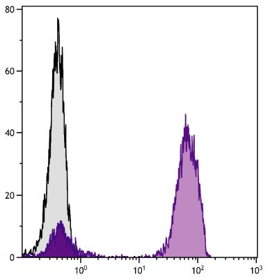
Human peripheral blood lymphocytes were stained with Mouse Anti-Human CD3-UNLB followed by Goat Anti-Mouse IgG1, Human ads-FITC (SB Cat. No. 1070-02). Human myofibroblasts were stained with anti-COX2 and anti-α-SMA followed by Goat Anti-Mouse IgG2a, Human ads-TXRD (SB Cat. No. 1080-07), Goat Anti-Mouse IgG1, Human ads-FITC (SB Cat. No. 1070-02), and DAPI. Image from Mattyasovszky SG, Hofmann A, B
Paraffin embedded human supraspinatus tendon tear section was stained with anti-CD206 followed by Goat Anti-Mouse IgG1, Human ads-FITC (SB Cat. No. 1070-02). Image from Goodier HC, Carr AJ, Snelling SJ, Roche L, Wheway K, Watkins B, et al. Comparis
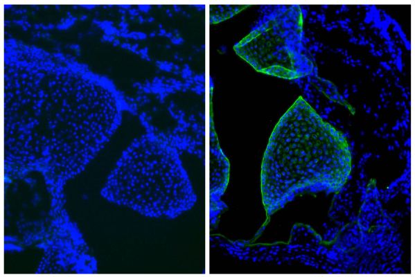
Frozen newborn mouse cartilage section was stained with Mouse IgG1-UNLB isotype control (SB Cat. No. 0102-01; left) and Mouse Anti-Type II Collagen-UNLB (right) followed by Goat Anti-Mouse IgG1, Human ads-FITC (SB Cat. No. 1070-02) and DAPI.
Contact