Anti-CD3 e (Ms,Hu,Mo,Ho,Bo,Sw,Rb,Dg,Ca,Mm,Ck,Bi,Fr) from Rat (HH3E) – unconj. for FFPE tissues
-
Overview
SKU DIA-303 Specificity Species Reactivity Bird, Bovine, Cat, Chicken, Dog, Frog, Horse, Human, Mammals, Monkey, Mouse, Rabbit, Swine
Immunogen Synthetic peptide from cytoplasmic epitope of CD3: ERPPPVPNPDYEPC
Host Species Isotype Clone Clonality (Mono-/Polyclonal) Application Immunofluorescence, Immunohistochemistry (frozen sections), Immunohistochemistry (IHC), Immunohistochemistry (Paraffin-embedded Sections), Western Blot
Conjugation Dilution Format 0.05% NaN3, 2% BSA, in PBS (pH 7.4), lyophilisate, purified antibody (from culture supernatant)
Product line / Topic Intended Use Temperature - Storage Temperature - Transport Search Code Manufacturer / Brand Uniprot_ID Gene_ID Alias CD3e, T-cell surface antigen T3/Leu-4 epsilon chain, T-cell surface glycoprotein CD3 epsilon chain, T3E
- Datasheets and Downloads
-
Additional Product Information
Reactivity:
Antibody clone HH3E has been validated specifically for the detection of murine CD3 in formalin-fixed paraffin-embedded tissue sections (mouse FFPE). It detects a conserved epitope on the CD3 epsilon chain in a broad variety of species.
Background:
CD3 is a defining feature of cells belonging to the T cell lineage and can therefore be used as T cell marker. Cluster of differentiation 3 (CD3) is composed of four distinct polypeptide chains CD3 gamma, CD3 delta, CD3 epsilon and CD3 zeta, that form a multimeric protein complex. The CD3 complex associates non-covalently with the T cell receptor (TCR) and serves as a T cell co-receptor. The CD3 components have long cytoplasmic tails that associate with cytoplasmic signal trans-duction molecules. The T cell antigen receptor (TCR) recognizes foreign antigens and translates such recognition events into in-tracellular signals that elicit a change in the cell from a dormant to an activated state. During T cell maturation the expression of CD3 migrates from the cytoplasm of pro-thymocytes to the cell-membrane of thymocytes. The specific appearance at all stages of T cell development make CD3 an ideal marker for normal T cells and T cell neoplasms (lymphomas, leukemias). Moreover, CD3 is a usefull immunohistochemical marker for T cells in tissue sections.
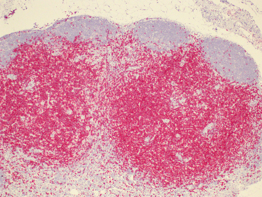
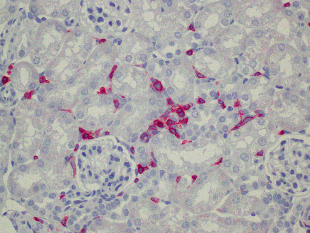
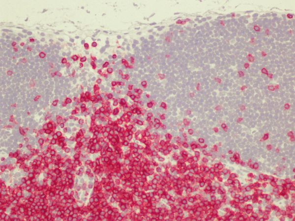
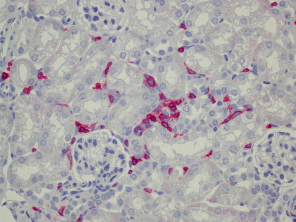
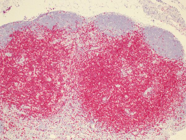
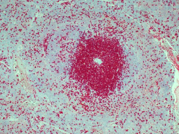
In a clinical setting in humans, CD3 serves as an important T cell marker for the classification of malignant lymphomas and leu-kemias. It can also be used to detect T cells in coeliac disease, lymphocytic and collagenous colitis. An anti-CD3 epsilon anti-body (Okt3) has been clinically approved for the induction of immunosuppression in organ transplantation. In animal studies anti-CD3 antibodies can induce tolerance to allografts.Immunohistochemistry of mouse CD3e (TCRE) in formalin-fixed paraffin-embedded tissue sections

A
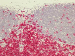
B
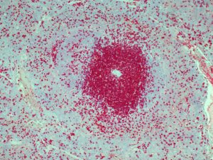
C
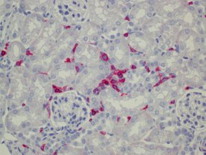
D
The monoclonal antibody clone HH3E specifically stains mouse tissue sections by IHC-FFPE: Lymph nodes (A,B), spleen (C) and kidney (D).
All sections were stained by an indirect alkaline phosphatase method according to standard procedures with antigen retrieval by high-temperature heating in citrate buffer and counterstaining with Haematoxylin.
Specific References:
- Hashimoto A, Sato T, Iyama S, Yoshida M, Ibata S, Tatekoshi A, Kamihara Y, Horiguchi H, Murase K, Kawano Y, Takada K, Miyanishi K, Kobune M, Ichimiya S, Kato J. Narrow-Band Ultraviolet B Phototherapy Ameliorates Acute Graft-Versus-Host Disease of the Intestine by Expansion of Regulatory T Cells. PLoS ONE. 2016;11(3):e0152823. PMCID: PMC4816442
General References:
- Leon F. Flow cytometry of intestinal intraepithelial lymphocytes in celiac disease. Journal of Immunological Methods 363: 177-186, 2011
- Smith-Garvin JE et al.. T cell activation. Annu Rev Immunol. 27:591-619, 2009
- Sapp H et al.The terminal ileum is affected in patients with lymphocytic or collagenous colitis. Am J Surg Pathol. 26(11):1484-1492, 2002
- Vernau W, Moore PF. An immunophenotypic study of canine leukemias and preliminary assessment of clon-ality by polymerase chain reaction. Vet Immunol Immunopathol. 69:145-164, 1999
- Mosnier et al. Lymphocytic and collagenous colitis: an immunohistochemical study. Am J Gastroenterol. 91(4):709-713, 1996
- Chetty R, Gatter K. CD3: Structure, function, and role of immunostaining in clinical practice. The Journal of Pathology 173(4): 303–307, 1994
- Salvadori S et al. Abnormal signal transduction by T cells of mice with parental tumors is not seen in mice bearing IL-2-secreting tumors. J Immunol. 153(11):5176-5182, 1994
- Nicolls MR et al. Induction of long-term specific tolerance to allografts in rats by therapy with an anti-CD3-like monoclonal antibody. Transplantation 55(3):459-468, 1993
Document-DownloadMSDS_dianova_V09Datasheet_DIA-303_V5
Quantity: 500 µl
Usual delivery time: 1 business day
345,00 € plus VAT
Free shipping inside Germany
QuantityRelated Products
DIA-310 – Anti-CD31 (Ms) from Rat (Clone: SZ31) for mouse FFPE tissue – 500 µl
DIA-450 – Anti-CD45R (Ms) from Rat (Clone: GHH45) for mouse FFPE tissue – 200 µl
-
Images
clone-HH3E-mouse-lymph-nodes-FFPE clone-HH3E-stains-mouse-kideny clone-HH3E-stains-mouse-lymph-nodes clone-HH3E-stains-mouse-spleen