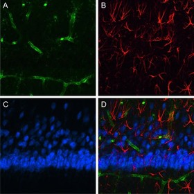
Selection of antibodies for simultaneous detection of more than one antigen depends on at least two important criteria:
A) Availability of secondary antibodies that do not recognize
1.) one another i.e. are derived from the same host species,
2.) other primary antibodies used in the assay system,
3.) immunoglobulins from other species present in the assay system, or
4.) endogenous immunoglobulins present in the tissues or cells under investigation.
B) Use of labels (fluorophores, enzyme-reaction products, electron-dense particles) that are well resolved.
The affinity-purified antibodies marked MinX (minimal cross-reactivity/reaction) have been specifically prepared to meet these criteria. They have been tested and/or adsorbed against IgG and/or serum proteins from other species.
Their cross-reactivity (X) with immunoglobulins of those indicated species is “minimal” (Min), which means below < 1%.
(Ha = Armenian Hamster, Hs = Syrian Hamster, Ck = Chicken, Hu = Human, Rb = Rabbit, Ms = Mouse, Gp = Guinea Pig, Ho = Horse, Rt = Rat, Bo = Bovine, Sh = Sheep, Sw = Swine, Go = Goat)
One of many possible multiple-labeling protocols using these reagents with minimal cross-reaction is shown in the following example. In this example, the secondary antibodies used in step 3 do not recognize each other since they are all made in donkey. They have been solid-phase adsorbed so that they do not recognize the other primary antibodies used in Step 2. Also, they do not react with endogenous rat immunoglobulins, which may be present in the rat tissue. However, the increase in specificity by adsorption to Ig/ serum proteins is at the expenses of the sensitivity: secondary antibodies from donkey, which are adsorbed against up to ten different species are less affine than secondary antibodies from goat, which have been adsorbed against Ig/ serum proteins from only three to five different species.
Note: Antibodies, which have been adsorbed against a closely related species, partly show a reduced epitope-recognition, so that monoclonal primary antibodies with rare IgG isotypes (IgG2b, IgG3), which are largely homologous to the species used for adsorption, may not be recognized as well any more.
Triple Labeling of Rat Tissue

* Also see: Example Fab blocking of tissue
Note: The detection of antigen A, B and C can be carried out simultaneously or consecutively. In a parallel approach, the antibodies in step 2 (b, d, f) and 3 (c, e, g) are applied as a cocktail, in the consecutive approach all single steps from a) to g) are done in successive incubations. In both approaches thorough washing between the antibody incubations and after blocking (a) is required. With heavy or persistent background in the consecutive approach, further blocking may be required before Steps d) and f). The multiple staining can be carried out in a parallel approach, if the staining and background characteristics of the antibodies after mixture are unchanged compared to single (or consecutive) stains with these antibodies. In mixtures, antibodies (immunoglobulins) may react with each other because of unspecific interactions and thereby may interfere with the antigen detection. For this reason no normal serum should be added to the antibody dilution buffer.
More Tips and Tricks:
>>> Selection of Fluorescent Dyes
>>> Multiple labelling with hapten-labelled primary antibodies>
For a review of multi-color immunofluorescence labeling with confocal microscopy see Brelje, Wessendorf, and Sorenson, “Multi-color laser scanning confocal immunofluorescence microscopy: Practical application and limitations.” In Cell Biological Applications of Confocal Microscopy, B. Matsumoto. Orlando, FL: Academic Press, Inc. (Methods Cell. Biol. 1993, Vol. 38, 97; Methods Cell. Biol. 2002, Vol. 70, 165).
Example protocol: Four-color Immunofluorescence Staining – Parallel Approach

Four-color staining of the Calcium binding proteins Calbindin, Parvalbumin, Calretinin, and the perineuronal net
(Ref.: Härtig et al. 1996, J. Neurosci. Methods 67: 89-95 and Schwaller et al. 1999, J. Neurosci. Methods 92: 137-144)
The Cytochemistry of calcium-binding proteins is often the method of choice for the imaging of morphological details of immune-positive nerve cells. Multiple labeling of these markers contributes to the understanding of complex structure-function relationships in the central nervous system.
Material: 30 µm, free floating, frozen sections of paraformaldehyde fixed rat brain.*
Note: The tissue sections are placed onto slides only after the completion of staining. All staining and washing steps are performed on free floating sections in wells of multi-well plates; sections are carefully transferred to wells containing washing or staining solution by means of an appropriate paintbrush.
All steps at room temperature:
- Wash tissue 3 x 10 min with 0,1 M Tris-buffered saline (TBS), pH 7.4
- Block unspecific binding sites in the tissue with 5% donkey normal serum (cat. 017-000-121) in TBS + 0,3% Triton X-100 (= ENS-TBS-T); 1 h
- Incubate with a cocktail of primary antibody and Wisteria floribunda agglutinin in ENS-TBS-T; 16 h* at room temperature:
• Mouse anti-Calbindin [1:200; Clone CL-300; Sigma]
• Rabbit anti-Parvalbumin [1:500; Swant]
• Goat anti-Calretinin [1:100; Swant]
• Biotinylated, reduced Wisteria floribunda Agglutinin (20 µg/ml; Sigma) - Wash sections 3 x 10 min with TBS
- Incubate with a cocktail of fluorescent secondary antibodies diluted in TBS (+ 2% bovine serum albumin, e. g. Fraction V)]; 1 h:
• Alexa Fluor 488 donkey anti-mouse IgG (cat. 715-545-151; 20 µg/ml)
• Cy3 donkey anti-rabbit IgG (711-165-152; 20 µg/ml)
• AMCA donkey anti-goat IgG (705-155-147; 30 µg/ml)
• Alexa Fluor 647-Streptavidin (016-600-084; 20 µg/ml) - Wash sections 3 x 10 min with TBS and with distilled or demineralized water (1 min)
- Draw up the sections onto fluorescent-free slides; let air dry
- Coverslip with e. g. Entellan (in Toluen; Merck) or with an aqueous mounting medium.
*Please note: This is not a standard protocoll. The incubation time for primary antibody depends on the properties of the antibody and the thickness of the tissue section and may vary from 1h to several hours at room temperature to several days at 4°C.
Depending on the availability of suitable primary antibodies, immunofluorescence detection can also be carried out on paraffin and cryostat sections as well as on wholemounts. For the sensitive visualization of certain, often membrane-associated antigens the use of detergents during histochemical procedures is important (commonly used: 0,1-1% Triton X-100 which is often added to the blocking solution for the primary antibodies).As the effect of Triton is irreversible, it only needs to be added once in the first incubation solution/s and its addition to secondary antibodies is not required. Higher Triton concentration in the incubation solutions used for different immunhistochemical procedures may cause artifacts, as for example myelin staining (see Weruaga et al. 1998).
