
Time saving, sensitive detection without any additional detection reagent (ECL) required
Anti-6His mouse monoclonal antibody clone 13/45/31-2 detects N- and C-terminally His-tagged proteines as well as internal His-tags with a high affinity.
The new HisDirect 60nm Gold conjugates detect His-tagged Protein in Western Blotting and Dot-Blot on nitrocellulose and PVDF membranes and can also be used in microbial colony and plate tests.
The unique HisDirect protocol with an incubation time of only 10 – 60 minutes is a one step protocol were no additional washing steps, detection reagent such as ECL or substrate incubation is required, making it a time and cost saving alternative to conventional blotting detection methods.

HisDirect is available as ready-to-use reagent and also as useful pads
| Ordering # | Quantity | Description |
|---|---|---|
| HisDirect-PA-010 | 10 Pads | HisDirect His-Tag WB Staining Pads (10x) pmol/fmol-Range (RUO) |
| HisDirect-PA-030 | 30 Pads | HisDirect His-Tag WB Staining Pads (30x) pmol/fmol-Range (RUO) |
| HisDirect-LP-010 | 10x10ml | HisDirect His-Tag WB Staining Solution pmol-Range ready-to-use (RUO) |
| HisDirect-LF-010 | 10x10ml | HisDirect His-Tag WB Staining Solution fmol-Range ready-to-use (RUO) |
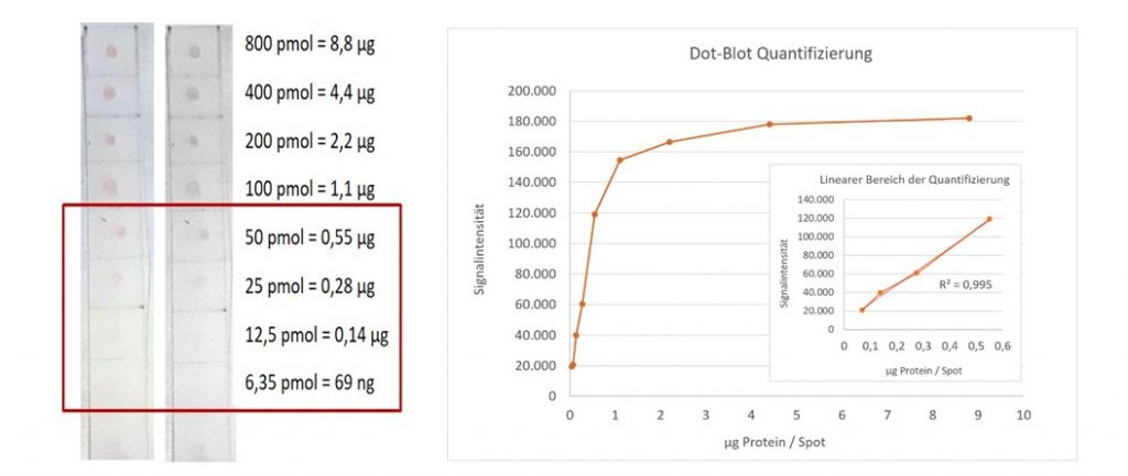
Quantification of DotBlots with HisDirect:
Additional anti 6His Antibody Formats and Resources: